missing translation for 'onlineSavingsMsg'
Learn More
Learn More
CD8b Monoclonal Antibody (eBioH35-17.2 (H35-17.2)), Super Bright™ 600, eBioscience™, Invitrogen™

Rat Monoclonal Antibody
161.00€ - 371.00€
Specifications
| Antigen | CD8b |
|---|---|
| Clone | eBioH35-17.2 (H35-17.2) |
| Applications | Flow Cytometry |
| Classification | Monoclonal |
| Conjugate | Super Bright 600 |
Description
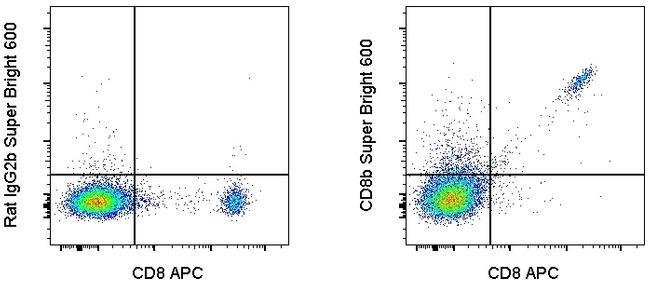
Description: The eBioH35-17.2 monoclonal antibody reacts with the mouse CD8 beta molecule. The CD8 beta chain associates with the CD8 alpha chain to form the CD8 alpha/beta heterodimer expressed on the surface of a majority of thymocytes, and on peripheral cytotoxic alpha beta TCR T cells. CD8 binds to MHC class I and plays a role in T cell development and activation of mature T cells. Applications Reported: This eBioH35-17.2 (H35-17.2) antibody has been reported for use in flow cytometric analysis. Applications Tested: This eBioH35-17.2 (H35-17.2) antibody has been tested by flow cytometric analysis of mouse splenocytes. This can be used at less than or equal to 0.25 μg per test. A test is defined as the amount (μg) of antibody that will stain a cell sample in a final volume of 100 μL. Cell number should be determined empirically but can range from 10^5 to 10^8 cells/test. It is recommended that the antibody be carefully titrated for optimal performance in the assay of interest. Super Bright 600 is a tandem dye that can be excited with the violet laser line (405 nm) and emits at 600 nm. We recommend using a 610/20 bandpass filter. Please make sure that your instrument is capable of detecting this fluorochrome. When using two or more Super Bright dye-conjugated antibodies in a staining panel, it is recommended to use Super Bright Staining Buffer (cat. SB-4400) to minimize any non-specific polymer interactions.
The CD8B antigen is a cell surface glycoprotein found on most cytotoxic T lymphocytes that mediates efficient cell-cell interactions within the immune system. The CD8 antigen, acting as a coreceptor, and the T-cell receptor on the T lymphocyte recognize antigens displayed by an antigen presenting cell (APC) in the context of class I MHC molecules. The functional coreceptor is either a homodimer composed of two alpha chains, or a heterodimer composed of one alpha and one beta chain. Both alpha and beta chains share significant homology to immunoglobulin variable light chains. This gene encodes the CD8 beta chain isoforms. Multiple alternatively spliced transcript variants encoding distinct membrane associated or secreted isoforms have been described. A pseudogene, also located on chromosome 2, has been identified.Specifications
| CD8b | |
| Flow Cytometry | |
| Super Bright 600 | |
| Rat | |
| Mouse | |
| 12526 | |
| IgG2b κ | |
| Affinity chromatography | |
| Antibody |
| eBioH35-17.2 (H35-17.2) | |
| Monoclonal | |
| Liquid | |
| RUO | |
| P10300 | |
| Cd8b1 | |
| Primary | |
| 4° C, store in dark, DO NOT FREEZE! | |
| Cd8b1 |
Spot an opportunity for improvement?Share a Content Correction
Product Content Correction
Your input is important to us. Please complete this form to provide feedback related to the content on this product.
Product Title