missing translation for 'onlineSavingsMsg'
Learn More

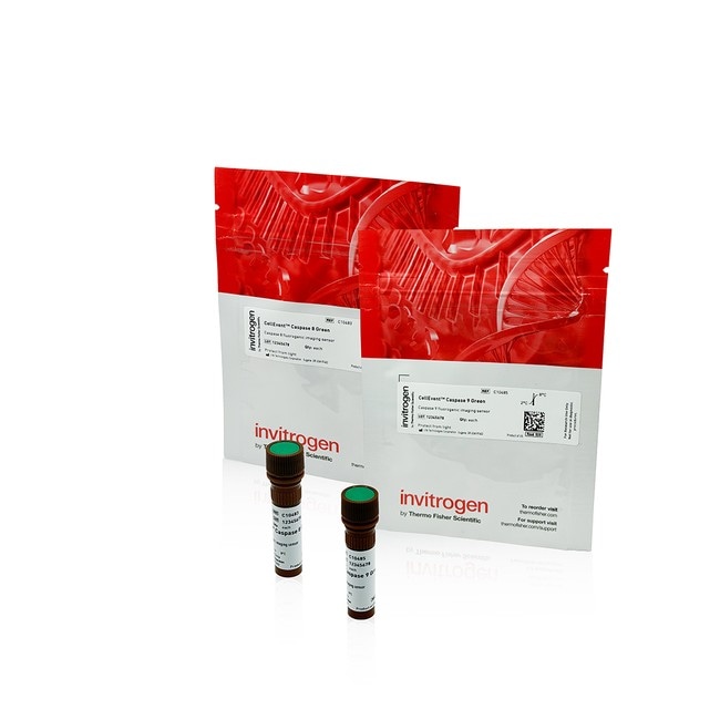
Invitrogen™ CellEvent Caspase 8 and 9 Detection Reagents

Product Code. 32096779
Change view
Click to view available options
Enzyme:
Caspase 8
Caspase 8 and Caspase 9
Caspase 9
Quantity:
1 x 300 μL Vial
2 x 300 μL Vials
5 x 300 μL Vials
Unit Size:
Each
5 product options available for selection
Product selection table with 5 available options. Use arrow keys to navigate and Enter or Space to select.
Product Code. Enzyme Quantity Unit Size
32096779 Caspase 9 5 x 300 μL Vials Each
32045443 Caspase 8 1 x 300 μL Vial Each
32096746 Caspase 8 5 x 300 μL Vials Each
32045278 Caspase 9 1 x 300 μL Vial Each
32096688 Caspase 8 and Caspase 9 2 x 300 μL Vials Each
Use arrow keys to navigate between rows. Press Enter or Space to select a product option. 5 options available.
5 options
This item is not returnable. View return policy
Product Code. 32096779 Supplier Invitrogen™ Supplier No. C10486
 Restricted Item
Estimated Shipment: 21-10-2026
Add to basket
Add to basket

missing translation for 'validMainframeMsg'

This item is not returnable. View return policy

CellEvent™ Caspase Detection Reagents are fluorogenic, cell-permeant substrates for detecting activated caspase-3/7, caspase-8, and caspase-9 enzymes in live cells. No-wash formats support endpoint, kinetic, high-content imaging, and microplate apoptosis assays.

CellEvent™ Caspase Detection Reagents are cell-permeant, fluorogenic substrates for detecting activated caspase-3/7, caspase-8, and caspase-9 during apoptosis. Following cleavage by the target caspase, the released dye binds DNA and produces bright nuclear fluorescence in apoptotic cells. The no-wash reagents support live-cell and fixed-cell fluorescence imaging, high-content analysis, and microplate reader-compatible assays. They can be used for endpoint, kinetic, continuous-culture, and multiplexed workflows in 2D or 3D cell models.

CellEvent Caspase-8 and CellEvent Caspase-9 Green Detection Reagents are cell-permeant, fluorogenic substrates for detecting activated initiator Caspases during apoptosis. Caspase-8 is generally activated downstream of death receptor signaling and Caspase-9 is generally activated through the mitochondrial apoptosome pathway. Measuring these enzymes can provide information about upstream extrinsic and intrinsic apoptosis signaling, while recognizing that pathway crosstalk and cell-specific responses can influence Caspase activity.

Product features

  • Detect activated Caspase-8 or Caspase-9 in living cells
  • Produce bright green nuclear fluorescence for image-based identification and segmentation of Caspase-positive cells
  • Use an add-and-read workflow with no cell lysis, permeabilization, or wash step required
  • Support endpoint, kinetic, continuous-culture, high-content imaging, and microplate workflows
  • Retain activated signal after formaldehyde-based fixation
  • Support multiplex analysis with spectrally compatible cell-health and viability probes

Substrate specifics and principles of action

Each reagent contains a four-amino acid peptide recognition sequence linked to a fluorogenic DNA-binding dye. CellEvent Caspase-8 Green Detection Reagent uses the IETD sequence, while CellEvent Caspase-9 Green Detection Reagent uses the LEHD sequence. In cells without activated target Caspase, the intact reagent remains nonfluorescent because the peptide prevents the dye from binding DNA.

Unlike other caspase detection reagents that bind a single dye molecule to an activated enzyme in an irreversible reaction, the fluorogenic induction of CellEvent dye by cleavage does not inhibit caspase enzymes in the cell enabling signal amplification as the dye binds DNA producing bright green nuclear fluorescence response in cells with Caspase activity.

The fluorescence can be detected by microscopy, automated high-content imaging, or a compatible microplate reader. Imaging helps provide single-cell context, while microplate readers provide a population-level measurement. Whole-well assays may require optimization of cell density, treatment conditions, and the proportion of Caspase-positive cells to generate sufficient signal.

CellEvent Caspase reagent formats and portfolio

The CellEvent portfolio also includes reagents for measuring downstream executioner Caspase activity. CellEvent Caspase-3/7 Red Detection Reagents (C10430, C10431) provide a red nuclear signal for experiments multiplexing with GFP or compatible green-fluorescent markers. CellEvent Caspase-3/7 Green ReadyProbes™ Reagent (R37111) is supplied in a ready-to-use dropper bottle for a simple labeling workflow. Other CellEvent Caspase-3/7 Green formats include C10723, C10423, C10432, and C10433.

Assay versatility for live-cell imaging and endpoint analysis

CellEvent Caspase-8 and Caspase-9 reagents can be added directly to living cells in complete culture medium or a compatible physiological buffer. For endpoint analysis, cells are typically incubated with reagent for 3060 minutes at 37°C before measurement. The green reagents have excitation and emission maxima of approximately 502/530 nm and can be detected using standard FITC or GFP optical configurations.

For kinetic and continuous-culture studies, the reagent can remain in complete medium during extended culture of 2D or 3D models. Time-lapse imaging with an environmentally controlled microscope or on-stage incubator can monitor the appearance, timing, and distribution of Caspase-positive nuclei during prolonged treatment. The workflow is compatible with systems such as the EVOS™ M7000 Imaging System equipped with the EVOS Onstage Incubator, or similarly configured microscopes with appropriate environmental control.

For microplate-based high-content imaging, the bright nuclear signal supports automated segmentation and measurements such as nuclear intensity or percentage of positive cells. The fluorescence response can also be measured with a microplate reader for population-level analysis.

The reagents must be activated by Caspases in living cells and cannot be applied to previously fixed samples to generate signal. After sufficient cleavage and signal development, cells may be preserved with a formaldehyde-based fixative for endpoint imaging, permeabilization, or subsequent analysis with compatible fixed-cell reagents.

Format advantages

  • Ready-to-dilute 100X aqueous concentrates at 500 μM
  • Direct dilution into compatible physiological buffer or complete culture medium
  • Storage at 28°C without preparation of a DMSO stock solution
  • Detection with standard FITC or GFP optical configurations

Applications

  • Drug discovery, compound profiling, and mechanism-of-action studies
  • Live-cell and time-lapse fluorescence imaging
  • On-stage incubator and continuous-culture experiments
  • High-content screening and automated image analysis
  • Microplate reader-based Caspase quantitation
  • Fixed-cell endpoint imaging after live-cell activation
  • Longitudinal studies in 2D and 3D cell models

Multiplex analysis of Caspase activity, mitochondrial function, and membrane integrity

TRUSTED_SUSTAINABILITY

Specifications

Quantity 5 x 300 μL Vials
Format Vial
Assay Apoptosis
Product Type Caspase detection reagent
Content And Storage •5 vials 300 μL 100x aqueous concentrate Caspase 9 detection reagent
•Store at 2°C to 8°C
•Protect from light
Compatible Cells Live mammalian cells
Excitation/Emission 502/530
No. of Reactions 100 Tests
Color Green
Incubation Time 30 to 60 min. or Continuous Culture
Product Line CellEvent
Detection Method Fluorescence
Formulation 500 μM 100x aqueous concentrate solution
For Use With (Application) Live cell imaging, 2D and 3D cell culture, continuous culture, drug discovery, apoptosis
Conjugate CellEvent Caspase 9 Green
For Use With (Equipment) Fluorescence Microscopes, High Content Instruments, Live Cell Imaging, Microplate Readers, Flow Cytometers
Enzyme Caspase 9
Show More Show Less
How many tests can I perform with each Invitrogen CellEvent Caspase-8 and Caspase-9 product?

The number of tests depends on the final assay volume. At 100 µL per well and 5 µM working solution, Invitrogen CellEvent Caspase-8 Green and Caspase-9 Green Detection Reagents yield approximately 300 tests per 300 µL vial. Practical yield may be lower because of pipetting loss, dead volume, and concentration optimization.

How should Invitrogen CellEvent Caspase-8 and Caspase-9 Detection Reagents be stored, and what formats are supplied?

Prepare the working staining solution fresh each day, protect the reagents from light, and avoid repeated freeze-thaw cycles. The shipped products are stable for six months from receipt when stored as directed. Invitrogen CellEvent Caspase-8 Green Detection Reagent, Cat. Nos. C10483 and C10484, and Invitrogen CellEvent Caspase-9 Green Detection Reagent, Cat. Nos. C10485 and C10486, are supplied as 100X, 500 µM aqueous concentrates stored at 2-8°C.

Can cleaved Invitrogen CellEvent Caspase-3/7 Detection Reagents stain cytoplasmic RNA?

Cytoplasmic RNA staining may occur after Invitrogen CellEvent Caspase Detection Reagents are cleaved. The intended assay readout is nuclear fluorescence from released dye binding to nucleic acids in caspase-positive cells.

How long can Invitrogen CellEvent Caspase Detection Reagents remain in continuous culture?

For continuous culture experiments the Invitrogen CellEvent Caspase Detection Reagents can be maintained in complete cell culture medium at its working concentration.

Can I use a nuclear counterstain with Invitrogen CellEvent Caspase Detection Reagents?

Yes, but add the nuclear counterstain after the Invitrogen CellEvent Caspase reagent signal has developed. For kinetic or continuous-culture experiments, add a Hoechst nuclear stain at the endpoint to reduce potential effects on normal cell physiology. Nuclear counterstain dyes may compete for nucleic acid binding, so optimize the combination.

Can Invitrogen CellEvent Caspase Detection Reagents be multiplexed with fluorescent antibodies or other probes?

Yes, when the labels are spectrally and chemically compatible. For live-cell surface labeling, apply and optimize the surface label before adding the CellEvent Caspase reagent. For intracellular immunofluorescence, allow the CellEvent Caspase reagent signal to develop in living cells before fixation, permeabilization, and antibody staining. Use single-color controls to evaluate spectral overlap, background, and staining sequence effects.

Can Invitrogen CellEvent Caspase-labeled cells be fixed, and can previously fixed cells be stained?

Invitrogen CellEvent Caspase-labeled living cells can be fixed after signal development, but previously fixed cells cannot be stained for caspase-dependent signal. Add CellEvent Caspase Detection Reagents to living cells before fixation. After signal development, cells may be fixed with 4% paraformaldehyde for 20 min at room temperature and washed three times as one example. Fluorescence may remain detectable after fixation, but retention depends on fixation, washing, storage, and imaging conditions. Image fixed samples promptly or verify the required storage interval. Wash steps may remove loosely attached apoptotic cells.

Can I combine Invitrogen CellEvent Caspase-3/7 Green Detection Reagent with PrestoBlue Cell Viability Reagent?

Yes. Invitrogen CellEvent Caspase-3/7 Green Detection Reagent has been demonstrated in combination with Invitrogen PrestoBlue Cell Viability Reagent in the same sample. CellEvent Caspase reagent fluorescence reports activated caspase, while PrestoBlue Cell Viability Reagent reports cellular metabolic activity as an indicator of viability. Follow the instructions for each reagent and use single-reagent controls to evaluate signal interference.

Can I quantify Invitrogen CellEvent Caspase Detection Reagents with a fluorescence microplate reader?

Yes. A compatible fluorescence microplate reader can measure Invitrogen CellEvent Caspase reagent fluorescence as a population-level whole-well response. Optimize cell density, treatment conditions, assay timing, and reader settings, and include untreated and induced controls. Use imaging when single-cell localization or percentage-positive measurements are required.

What controls should I include in an Invitrogen CellEvent Caspase experiment?

Include untreated or vehicle-treated cells, an induced positive control, a no-reagent background control, and an inhibitor-pretreated condition when appropriate. The source protocol lists camptothecin with Z-DEVD-FMK for caspase-3/7 and staurosporine with Z-IETD-FMK or Z-LEHD-FMK for caspase-8 or caspase-9. Optimize inducer and inhibitor concentrations and timing for the cell model.

How can I quantify Invitrogen CellEvent Caspase activity by high-content imaging?

Quantify nuclear fluorescence using measurements such as percentage of caspase-positive cells, nuclear fluorescence intensity, and nuclear morphology. Use consistent acquisition settings and define positive thresholds relative to untreated controls.

Which fluorescence filters should I use with Invitrogen CellEvent Caspase Detection Reagents?

CellEvent Caspase-3/7 Green, Caspase-8 Green, and Caspase-9 Green Detection Reagents, which have approximate excitation/emission maxima of 502/530 nm. Use a Texas Red dye optical configuration for CellEvent Caspase-3/7 Red Detection Reagent, which has approximate maxima of 590/610 nm.

Can Invitrogen CellEvent Caspase Detection Reagents be used with suspension, primary, or stem cells?

Yes. The protocol can be adapted to additional cell types, but different cell models will require optimization. The user guide protocol was optimized using HeLa, A673, and U-2 OS cells. When evaluating a new cell model, titrate the reagent, verify induction conditions, and include positive, negative, and inhibitor controls.

Can Invitrogen CellEvent Caspase Detection Reagents be used with both 2D and 3D cell models?

Yes. Invitrogen CellEvent Caspase Detection Reagents may remain in complete medium during extended culture of planar 2D and 3D models. For spheroids, organoids, or other thick models, optimize reagent working concentration, treatment conditions, imaging depth, and analysis because geometry and diffusion can affect the measured response.

Can Invitrogen CellEvent Caspase Detection Reagents be used for time-lapse imaging with an on-stage incubator?

Yes. Use an environmentally controlled microscope or on-stage incubator maintained at the required culture conditions such as the Invitrogen EVOS M7000 Imaging System with the Invitrogen EVOS Onstage Incubator.

Do Invitrogen CellEvent Caspase Detection Reagents require washing or medium replacement?

No. Invitrogen CellEvent Caspase Detection Reagents are designed for add-and-read use in complete culture medium or compatible physiological buffer. Medium composition and cell type can affect performance, so optimize reagent concentration and acquisition settings for new models. Avoiding a wash step may also reduce loss of loosely attached apoptotic or dying cells.

When should Invitrogen CellEvent Caspase fluorescence appear?

Signal timing depends mainly on when the target caspase becomes active. For endpoint measurements, allow 30-60 minutes at 37°C after adding the reagent before measurement.

What working concentration should I use for Invitrogen CellEvent Caspase Detection Reagents?

Start at 5 µM for Invitrogen CellEvent Caspase-3/7 Green, Caspase-3/7 Red, Caspase-8 Green, and Caspase-9 Green Detection Reagents; optimize within 2-10 µM for a new model. For CellEvent Caspase-3/7 Green ReadyProbes Reagent, add two 40 µL drops per mL of complete medium.

How do I prepare and run an endpoint Invitrogen CellEvent Caspase assay?

Add a freshly prepared 10X Invitrogen CellEvent Caspase staining solution to living cells at a 1:10 dilution in complete culture medium or a compatible physiological buffer. Treat the cells with the selected apoptosis inducer as required, incubate with the reagent for 30-60 minutes at 37°C, and measure fluorescence. Cell lysis, permeabilization, and washing are not required before measurement.

Can I measure caspase-8, caspase-9, and caspase-3/7 in the same experiment?

Yes, but use matched parallel wells for Invitrogen CellEvent Caspase-3/7 Green, Caspase-8 Green, and Caspase-9 Green Detection Reagents since their spectra overlap, so they are not distinguishable in the same fluorescence channel when combined. CellEvent Caspase-3/7 Red Detection Reagent may be paired with either green reagent after experimental verification with single-color controls.

Why can different mammalian cell types show different caspase responses?

Caspase responses can vary because cell type, culture medium, cell density, treatment, pathway composition, and activation timing affect staining and signal. Evaluate new cell models with untreated, induced, and inhibitor-treated controls.

When should I use Annexin V instead of an Invitrogen CellEvent Caspase Detection Reagent?

Choose Annexin V to detect phosphatidylserine exposure at the plasma membrane, and choose Invitrogen CellEvent Caspase Detection Reagents to detect activated caspases and nuclear fluorescence. The methods measure different apoptotic events and should not be treated as interchangeable.

When should I choose an Invitrogen CellEvent Caspase assay instead of a luminescence-based caspase assay?

Choose Invitrogen CellEvent Caspase Detection Reagents when you need live-cell monitoring, kinetic measurements, image-based cellular context, or signal that can be retained after fixation. Luminescence-based caspase assays commonly lyse cells and generate a population-level well signal for plate-based endpoint quantitation. Invitrogen CellEvent Caspase assays can also be measured with compatible fluorescence microplate readers.

How are Invitrogen CellEvent Caspase Detection Reagents different from FLICA reagents?

Invitrogen CellEvent Caspase reagents are cleavable substrates, whereas FLICA reagents are fluorescent caspase inhibitors that covalently bind active caspases. FLICA workflows typically require removal of unbound reagent, and the bound reagent inhibits the labeled enzyme. CellEvent Caspase reagents generate nuclear fluorescence without inhibiting caspase activity and do not require a wash step.

How should I interpret different caspase-8, caspase-9, and caspase-3/7 activation patterns?

Interpret the signals as measurements of caspase activity, not definitive evidence of a single apoptosis pathway. Pathway crosstalk, feedback, stimulus, cell type, and activation timing can affect which caspases become active. Compare matched cell density, treatment conditions, acquisition settings, and time points when evaluating activation patterns.

Which Invitrogen CellEvent Caspase Detection Reagent should I choose?

Choose according to the caspase target and fluorescence channel. Use Invitrogen CellEvent Caspase-3/7 Green or Red Detection Reagent for downstream executioner caspase activity, Invitrogen CellEvent Caspase-8 Green Detection Reagent for initiator activity generally associated with death-receptor signaling, and Invitrogen CellEvent Caspase-9 Green Detection Reagent for initiator activity generally associated with mitochondrial apoptosome signaling. Choose the red Caspase-3/7 reagent when GFP or another green fluorescent marker is present.

How do Invitrogen CellEvent Caspase Detection Reagents generate fluorescence, and which recognition sequences do they use?

Each Invitrogen CellEvent Caspase Detection Reagent contains a caspase-recognition peptide linked to a proprietary fluorogenic DNA-binding dye. Caspase cleavage releases the dye so it can bind nuclear DNA and fluoresce. CellEvent Caspase-3/7 reagents use the DEVD recognition sequence, CellEvent Caspase-8 Green Detection Reagent uses IETD, and CellEvent Caspase-9 Green Detection Reagent uses LEHD.

What are Invitrogen CellEvent Caspase Detection Reagents?

Invitrogen CellEvent Caspase Detection Reagents are cell-permeant, fluorogenic substrates used to detect activated caspase-3/7, caspase-8, or caspase-9 in living cells. They generate nuclear fluorescence in caspase-positive cells and support endpoint, kinetic, continuous-culture, high-content imaging, and fluorescence microplate reader workflows.


For Research Use Only. Not for use in diagnostic procedures.

Product Title
Select an issue

By clicking Submit, you acknowledge that you may be contacted by Fisher Scientific in regards to the feedback you have provided in this form. We will not share your information for any other purposes. All contact information provided shall also be maintained in accordance with our Privacy Policy.